大脳皮質は、様々な情報を処理する。
magnetic resonance imaging (MRI) で解析される、ヒトの大脳皮質の表層や厚さは、神経学的、精神学的、行動学的な機能と相関する事が知られている。
けれども、遺伝子多型が大脳皮質の構造に影響するかどうか、今まで明らかではなかった。
今回、オーストリア、アメリカ、オランダ etc.の研究グループは、遺伝子多型と脳皮質の構造の違いについて調べた。
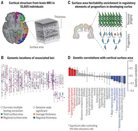
The human cerebral cortex is important for cognition, and it is of interest to see how genetic variants affect its structure. Grasby et al. combined genetic data with brain magnetic resonance imaging from more than 50,000 people to generate a genome-wide analysis of how human genetic variation influences human cortical surface area and thickness. From this analysis, they identified variants associated with cortical structure, some of which affect signaling and gene expression. They observed overlap between genetic loci affecting cortical structure, brain development, and neuropsychiatric disease, and the correlation between these phenotypes is of interest for further study. Science , this issue p. [eaay6690][1] ### INTRODUCTION The cerebral cortex underlies our complex cognitive capabilities. Variations in human cortical surface area and thickness are associated with neurological, psychological, and behavioral traits and can be measured in vivo by magnetic resonance imaging (MRI). Studies in model organisms have identified genes that influence cortical structure, but little is known about common genetic variants that affect human cortical structure. ### RATIONALE To identify genetic variants associated with human cortical structure at both global and regional levels, we conducted a genome-wide association meta-analysis of brain MRI data from 51,665 individuals across 60 cohorts. We analyzed the surface area and average thickness of the whole cortex and 34 cortical regions with known functional specializations. ### RESULTS We identified 306 nominally genome-wide significant loci ( P Identifying genetic influences on human cortical structure. ( A ) Measurement of cortical surface area and thickness from MRI. ( B ) Genomic locations of common genetic variants that influence global and regional cortical structure. ( C ) Our results support the radial unit hypothesis that the expansion of cortical surface area is driven by proliferating neural progenitor cells. ( D ) Cortical surface area shows genetic correlation with psychiatric and cognitive traits. Error bars indicate SE. IMAGE CREDITS: (A) K. COURTNEY; (C) M. R. GLASS The cerebral cortex underlies our complex cognitive capabilities, yet little is known about the specific genetic loci that influence human cortical structure. To identify genetic variants that affect cortical structure, we conducted a genome-wide association meta-analysis of brain magnetic resonance imaging data from 51,665 individuals. We analyzed the surface area and average thickness of the whole cortex and 34 regions with known functional specializations. We identified 199 significant loci and found significant enrichment for loci influencing total surface area within regulatory elements that are active during prenatal cortical development, supporting the radial unit hypothesis. Loci that affect regional surface area cluster near genes in Wnt signaling pathways, which influence progenitor expansion and areal identity. Variation in cortical structure is genetically correlated with cognitive function, Parkinson’s disease, insomnia, depression, neuroticism, and attention deficit hyperactivity disorder. [1]: /lookup/doi/10.1126/science.aay6690 [2]: pending:yes
遺伝子による脳の構造の違い
著者らは、60コホート、51,665人のgenome-wide association meta-analysis (GWAS) のデータと脳MRIのデータを検証し、34の皮質野で脳表層や皮質の厚さを調べた。
結果、33,992人の被験者で、306の遺伝子多型が脳皮質の構造に違いを持つ事が分かった。
脳表層の面積は、脳の発達段階で神経前駆細胞の活動を調節する遺伝子に影響を受けていると考えられる。
一方、脳皮質の厚さは、胎生中期以降のプロセス、例えば髄鞘形成、分岐やシナプスの刈り込みなどの変化を反映していると考えられる。
175の遺伝子多型が脳の一部のエリアの表層面積に、10の遺伝子多型が部分的な皮質の厚さに影響しているようだった。
さらに、これらの被験者の認知機能や教育歴等との相関も検証した。
パーキンソン病(Parkinson's disease, PD)の患者は、全体の脳表層の面積が大きい傾向があった。
一方、不眠、注意欠陥多動性障害(attention deficit hyperactivity disorder, ADHD)、鬱症候群、うつ病、神経症などの患者では、全体の脳表層の面積が小さい傾向があった。
My View
以前、殺人などの重大な犯罪を犯す人は、その一歩手前で留まる人と比べて脳の灰白質のボリュームが少ない、という報告がありました(Sajous-Turner et al. Brain Imagina Behav, 2019)が、不眠やADHD、神経症の方でも皮質の表面性が減っている事があるようです。
病気以外でも、脳の構造がちょっとずつ違う事で、ストレスに弱かったり、なかなか集中できなかったり、人とうまく付き合えなかったり等、その人の個性のような形で表れるのかもしれません。
それにしてもこのプロジェクト、共著者&コレスポの多い事……
References
- Grasby et al., The genetic architecture of the huma cerebral cortex.Science 20 Mar 2020: Vol. 367, Issue 6484, eaay6690 doi: 10.1126/science.aay6690
- University of North Carolina Health Care. "Worldwide scientific collaboration unveils genetic architecture of gray matter." ScienceDaily. ScienceDaily, 26 March 2020. <www.sciencedaily.com/releases/2020/03/200326160801.htm>.
- Sajous-Turner, A., Anderson, N.E., Widdows, M. et al. Aberrant brain gray matter in murderers. Brain Imaging and Behavior (2019). https://doi.org/10.1007/s11682-019-00155-y

